Tag Archives: orthopedics news

The price of delirium: New study finds nearly half of patients have delirium

OrthopedicsDelirium associated with 7.4 additional hospital days and $8,000 more in hospital costs

A new study presented at the 2015 Annual Meeting of the American Academy of Orthopaedic Surgeons (AAOS) found that 48 percent of hip fracture patients, age 65 and older, had delirium, or acute confusion, before, during and after surgery (perioperative), resulting in significantly longer hospital stays and higher costs for care.Approximately 300,000 Americans are hospitalized with hip fractures each year. The risk is particularly high in post-menopausal women who face an increased risk for osteoporosis, a disease that diminishes bone mass and increases fracture risk. Delirium is common among older hip fracture patients, and multiple studies have found that patients with postoperative delirium are more likely to have complications, including infections, and less likely to return to their pre-injury level of function. Delirium patients also are more frequently placed in nursing homes following surgery, and have an increased rate of mortality.


Read the rest of the article http://www.medicalnewstoday.com/releases/291377.php.

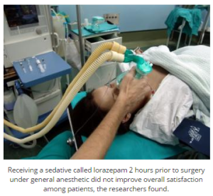
Patients may not benefit from sedatives prior to surgery, study suggests

ALL Surgery_Plastic_Orthopedic_Cosmetic_Neurosurgery_ETCPrior to undergoing a surgical procedure, many patients are prescribed sedatives to help reduce stress and anxiety before and after surgery. But a new study published inJAMA has questioned the benefits of these sedatives after finding they may not improve patients’ experience and may even do more harm than good.Benzodiazepines are a class of drugs commonly used to help reduce a patient’s perioperative stress or anxiety – the stress and anxiety experienced before and after surgery.However, the study researchers – including Dr. Axel Maurice-Szamburski of the Hôpital de la Timone Adulte in Marseille, France – note that benzodiazepines have been associated with a number of side effects, including drowsiness, insomnia and cognitive impairment.While benzodiazepines are deemed effective for reducing anxiety, the researchers note there is insufficient evidence to conclude their use leads to a better perioperative experience for patients.
Read the rest of the article http://www.medicalnewstoday.com/articles/290202.php.

New study finds same patient mortality rates for experienced and new surgeons

Thoracic Surgery_Orthopedic Surgery_Neurosurgery_All Surgery EXCEPT PlasticAdditional study needed to affirm consistency of findings to latest surgeon training practices, other factors, say researchersThere is no statistical difference between the patient mortality rates of new and experienced surgeons a study using a newly developed statistical methodology and conducted by a research team comprised of medical doctors and statisticians has found.Because surgical training was radically changed in recent years–including a reduction of six to 12 months of training time–and other factors, the research team said further study will be needed to ensure the findings generalize.Researchers involved in this study are Dr. Rachel R. Kelz, associate professor of surgery; Dr. Jeffrey Silber, professor of pediatrics, anesthesiology and critical care; Paul Rosenbaum, professor of statistics; and Sam Pimentel, a doctoral student in statistics–all of the University of Pennsylvania.

Read the rest of the article at http://www.medicalnewstoday.com/releases/289867.php

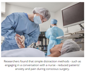
Distraction techniques may reduce pain, anxiety during conscious surgery

Emergenc Medicine_Surgery_Anesthesiology_PsychiatryFor some surgical procedures – such as the removal of varicose veins – the patient remains awake. Though safe, the patient can experience some pain and anxiety. But in a new study, researchers from the University of Surrey in the UK say simple methods of distraction could help ease such experiences.To reach their findings, published in the European Journal of Pain, Prof. Jane Ogden and colleagues enrolled 398 patients who were due to undergo varicose veinsurgery.For this type of surgery, patients typically remain awake, receiving only a local anesthetic.The researchers note that previously, patients have reported unfamiliar feelings, sounds and smells during the procedure. Some have also reported feeling a burning sensation, while others have said listening to conversations about the procedure between the surgeon and nurse makes them feel uneasy. Patients have also reported feeling anxious during the surgery.

Read the rest of the article at   http://www.medicalnewstoday.com/articles/288755.php.

The Royal College of Anaesthetists calls for joined up care for high risk surgical patients

Anesthesiology_Plastic Surgery_Orthopedic Surgery_Thoracic Surgery-Neurosurgery_Emergency Medicine


The Royal College of Anaesthetists (RCoA) shared its vision for the future of perioperative healthcare provision across the UK. Speaking at a stakeholder event, President of the RCoA Dr J-P van Besouw said: “With more than ten million patients undergoing surgery each year in the NHS, approximately 15% of whom are deemed to be high-risk, a care pathway that can harness multi-disciplinary working, reduce variation and improve patient outcomes must be our goal.”

Health experts, including Celia Ingham Clark, Director for Reducing Premature Mortality, NHS England, highlighted the scale of the unmet need for the high-risk surgical patient and the future principles for a more efficient and effective care pathway, at the event attended by a cross section of health bodies.

J-P van Besouw said: “Lowering the rate of avoidable harm – associated with many preventable complications and deaths – will make a significant saving to the NHS annual surgical bill, which at present runs at around GBP16 billion. The College believes that improved perioperative care is an efficient and effective solution.”

The RCoA proposed that care pathways designed around the individual needs of the complex surgical patient and delivered by a multi-disciplinary perioperative care team would mean that patients should receive the most appropriate care possible. Anaesthetists are uniquely positioned to lead such teams and to ensure a better continuum of care for patients, before, during and after surgery. Such an integrated approach should minimise inappropriate care, reduce cancellations, complications and readmissions and ensure quicker recovery and discharge from hospital. Fundamentally patients should receive better quality care, and experience better outcomes at a lower cost.

As well as improved patient centered care, the economic case for change is an important factor to be considered. The NHS Five Year Forward view states that by 2020/21, there could be a mismatch between resources and patient needs of nearly GBP30 billion a year. Demand, efficiency and funding are the key considerations and the evidence would indicate that perioperative medicine would be a cost effective solution through the reduced costs of treating complications.

In driving this agenda the College stated that it intended to work collaboratively with stakeholders in primary care, other colleges, specialties and workforce planners in Health Education England (HEE) and the devolved nations, to develop and commission new standards of patient care, embed continuous data driven quality improvement into the care of surgical patients, and explore solutions to create an appropriate workforce for a sustainable future.

“We do not underestimate the challenges that we face in delivering this agenda,” said J-P van Besouw. “We are asking colleagues, policy makers and commissioners to share this vision and to use the resources that we have developed, including a short animated film [http://www.rcoa.ac.uk/periopmed/animation], to start the conversation on what perioperative practice should look like and to work with us to develop integrated pathways of care, based on the many existing examples of excellent perioperative practice from across the UK.”

Adapted by MNT from original media release

http://www.medicalnewstoday.com/releases/288455.php


An alternative for pain control after knee replacement surgery

Orthopedics_Anesthesiology

It’s estimated that more than half of adults in the United States diagnosed with knee osteoarthritis will undergo knee replacement surgery. While improvements in implantable devices and surgical technique has made the procedure highly effective, pain control after surgery remains a common but persistent side effect for patients.

A Henry Ford Hospital study, presented recently at the American Association of Hip and Knee Surgeons meeting in Dallas, found that injecting a newer long-acting numbing medicine called liposomal bupivacaine into the tissue surrounding the knee during surgery may provide a faster recovery and higher patient satisfaction.

“The pain scores for this injection technique averaged about 3/10, which is similar to the pain scores seen with our traditional method,” says Jason Davis, M.D., a Henry Ford West Bloomfield Hospital joint replacement surgeon and the study’s senior author. “Patients had pain relief for up to two days after surgery and better knee function compared with the traditional method.”

It is estimated that the number of total knee replacement surgeries has more than tripled from 1993 to 2009. Arthritisis the most common cause of chronic knee pain and disability. However, a June 2014 study found that 95 percent of knee surgeries are attributed to the epidemic of overweight and obesity in the United States.

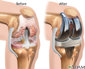
During the two-hour knee replacement procedure, the orthopedic surgeon removes the damaged cartilage and bone, and inserts a knee implant to restore the alignment and function of the knee. More than 90 percent of knee replacements are functioning 15 years after surgery, according to the American Academy of Orthopaedic Surgeons.

In the Henry Ford study, 216 patients were evaluated for pain control the first two days after surgery from October 2012 to September 2013. Half of the patients received the traditional pain control method with continuous femoral nerve blockade, in which common numbing medicine is injected into the groin area, blunting the main nerve down the front of the knee. This method uses a pain pump to extend pain control for two days but causes some leg weakness. “Pain control came at the price of weakness and made patients somewhat tentative when walking during their hospital stay,” Dr. Davis says.

The other half of patients received the liposomal bupivacaine injection at the site of the surgery.

Dr. Davis says many patients were able to walk comfortably within hours after surgery.

Dr. Davis says the injection around the knee itself “optimizes pain control early on” without the side effects of the traditional technique. “Function-wise, it was a lot easier for patients to move around more confidently,” he says. “In the past decade, we’ve made major advancements in pain control for knee replacement surgery. This option is a promising, viable one for our patients.”

Adapted by MNT from original media release

http://www.medicalnewstoday.com/releases/287423.php


The difficulties of treating shoulder pain in baseball pitchers

Anesthesiology

Results of treating shoulder pain in baseball pitchers and other throwing athletes are not as predictable as doctors, patients and coaches would like to think, according to a report in the journal Physical Medicine and Rehabilitation Clinics of North America.

Nickolas Garbis, MD, an orthopedic surgeon who specializes in shoulder and elbow injuries at Loyola University Medical Center, is the primary author.

Shoulder pain occurs in athletes who play sports that require rapid acceleration and deceleration of the throwing arm. They include baseball pitchers, tennis players, softball pitchers and javelin throwers, as well as athletes who play handball and water polo.

Overhead throwing generates a large amount of stress on the shoulder, which is one of the most mobile joints in the body. This makes it vulnerable to injury.

It is difficult to diagnose the cause of shoulder pain. The shoulder is comprised of four joints, and a problem with any of them can cause pain and affect performance. Moreover, many of these structures are deep in the shoulder and therefore difficult to examine by touch. Also, the same kind of pain can be due to multiple causes. For example, pain in the front of the shoulder can be due to rotator cuff tendinitis, rotator cuff tears, biceps tendinitis, shoulder instability, shoulder stiffness and several other causes.

“A systemic approach, and some experience, can help the clinician become more familiar with which constellation of findings in these athletes is not normal,” Dr. Garbis and co-author Edward McFarland, MD, write.

Shoulder problems can begin during adolescence. Little League shoulder, an injury to the growth plate in the shoulder, is one of the most common. Adolescent pitchers most at risk for injuries are those who compete on traveling teams. Overuse injuries can lead to more serious mechanical injuries. Adhering to pitch counts should reduce injuries and decrease fatigue.

Treatment should be primarily nonsurgical. Nonsurgical options include icing the shoulder and judicial use of nonsteroidal anti-inflammatory medications such as ibuprofen and naproxen. Rehabilitation can restore a normal muscular balance. Rest can help, but it should not be prolonged, because the shoulder could become deconditioned.

If nonsurgical options fail, arthroscopic surgery can be considered. For example, surgical repair or trimming of partial rotator cuff tears can be highly successful, returning as many as 89 percent of college and professional pitchers back to play. However, the type of surgery needed depends upon the patient’s shoulder problem.

http://www.medicalnewstoday.com/releases/286793.php


Pain relief for kids in the ER without a needle

Emergency Medicine_Pediatrics_Orthopedic Surgery_Anesthesiology

Children in emergency departments can safely be treated for pain from limb injuries using intranasal ketamine, a drug more typically used for sedation, according to the results of the first randomized, controlled trial comparing intranasal analgesics in children in the emergency department. The study was published online recently in Annals of Emergency Medicine (“The PICHFORK (Pain in Children Fentanyl OR Ketamine) Trial: A Randomized Controlled Trial Comparing Intranasal Ketamine and Fentanyl for the Relief of Moderate to Severe Pain in Children with Limb Injuries”).

“This is great news for emergency physicians and their young patients, especially those who may not tolerate other intranasal pain medications such as fentanyl,” said lead study author Professor Andis Graudins, MD, of Monash University in Clayton, Victoria, Australia. “For children in pain and distress, the option of treating their pain without a needle is a huge benefit as well. The intranasal option using fentanyl is accepted already for children, but the safe use of ketamine is new.”

Researchers compared pain relief resulting from ketamine and fentanyl, both delivered intranasally, for children 3 to 13 years old whose pain from isolated limb injuries registered seven or higher on a 10 point scale. Median baseline pain rating was eight out of ten. After 30 minutes, the median reductions in pain for ketamine were 4.45 and for fentanyl were 4.0. The pain reduction was maintained in both groups at 60 minutes. Satisfaction for ketamine was slightly higher at 83 percent. Fentanyl had a 72 percent satisfaction rating.

Adverse events were reported more frequently for ketamine (78 percent of patient) than for fentanyl (40 percent of patients), but they were all mild (dizziness or drowsiness were common).

“Ketamine is a great alternative for injured children in the ER who may not be able to tolerate opiates, like fentanyl,” said Prof. Graudins. “And being able to deliver pain-relief with minimal upset, such as that triggered in some children by even the sight of needles, is a great boon to our youngest patients.”

http://www.medicalnewstoday.com/releases/286526.php


Laboratory breakthrough offers promise for spinal cord injury patients to breathe on their own again

Orthopedic Surgery

Case Western Reserve researchers have developed a procedure that restores function to muscles involved in the control of breathing – even when they have been paralyzed for more than a year. The breakthrough offers hope that one day patients with severe spinal cord injuries will be able to breathe again without the assistance of a ventilator.

Principal investigator Philippa M. Warren, PhD, presented the results Nov. 17 at Neuroscience 2014, the annual meeting of the Society for Neuroscience. The research represents a critical step forward in efforts to reverse even long-term paralysis of muscles within the diaphragm that are activated by nerve fibers that extend from the upper part of the brain stem. When those fibers are damaged in the spinal cord, electrical signals from the brain cannot reach motor nerves that leave the spinal cord to activate muscles that control vital functions. This new research offers a two-step approach to repair the part of the damage that blocks those signals.

“We show that respiratory paralysis can be reversed at long intervals after spinal cord injury,” said Warren, aneurosciences researcher at MetroHealth Medical Center, which is affiliated with Case Western Reserve University School of Medicine. “This has the potential to alleviate the long suffering of currently injured patients, improving their quality, and potentially length, of life.”

Investigators focused their research on a group of nerves that extend from the respiratory control center in the brain stem down to the C3 through C5 vertebral levels of the spinal cord located in the middle of the neck. These fibers, or brain axons, control the diaphragm muscle in its critical function of breathing. Any injury to the spinal cord above the C3 vertebra can cause widespread muscle paralysis leading to difficulties in breathing, but also moving, regulation of cardiac output, and sexual function. Unfortunately, these injuries high in the neck are the most common among sufferers of spinal cord trauma.

Following injury to the spinal cord, damaged nerve fibers die, causing loss of the connections between the brain and muscles of the body. To help preserve tissue immediately after injury, a scar forms at the site of the trauma and extends the distance of several inches up and down the spinal cord. This scar tissue is very dense, contains sugars that inhibit new neuronal growth, and does not reduce in length or intensity over time. The consequence is that new connections cannot form to enable muscle function after injury, which is exceptionally important to breathing.

Spinal cord injury-induced paralysis of the respiratory muscles causes low oxygen in the blood, increases the body’s drive to breathe and drives any functioning respiratory muscles to work harder. The breathing capacity of the spinal cord-injured is often not enough to fully support a patient’s life. However, if new nerve fibers or connections can form in the spinal cord, then pathways can be activated to restore respiratory function. So Case Western Reserve researchers devised a technique to treat the injury site with a specially designed enzyme to reopen connections and to apply respiratory therapy to strengthen the remaining functioning respiratory muscles.

In laboratory animals, investigators used the combination technique to restore respiratory function many months after the injury. First, they injected the chondroitinase enzyme at the site of respiratory nerves in the spinal cord to remove the inhibiting sugars from scar tissue. The action of the enzyme enabled both the formation of new connections and stimulation of latent pathways in the respiratory motor system. Second, the animals were exposed to brief periods of conditions with low oxygen, making them breathe harder and faster to rehabilitate the respiratory muscles. This treatment approach is referred to as intermittent hypoxia.

The combination enzyme injection and intermittent hypoxia treatment boosts levels of serotonin. Commonly known to help relieve anxiety disorders, serotonin also acts more broadly as a neurotransmitter to help stimulate nerve cells. By increasing serotonin at nerve connections and at the specific receptors on the fibers themselves, the researchers were able to help restore diaphragm function back to normal levels in the animals. This finding is extraordinary not only because function to the paralyzed muscle was completely restored, but also because researchers were able to achieve breathing in animals that had been injured for a year and a half.

“It is remarkable to reactivate the diaphragm and breathing in a chronically injured animal that has had a paralyzed half diaphragm most of its life,” said Jerry Silver, PhD, a Case Western Reserve professor of neurosciences who collaborated in the research.

While these results are encouraging, more research is required to perfect the treatment. More than two-thirds of the animals in the study responded to the combined treatment strategy, while the treatment had no effect on the remaining animals. Two thirds of the animals that responded to the combined treatment resumed normal breathing, while the other third experienced erratic breathing in the injured muscle.

Investigators found that the animals with erratic breathing were flooded with too much serotonin during their treatment. A simple fix involved administering a serotonin receptor blocker, which restored these animals to normal breathing. Researchers are currently studying further the serotonin-overload phenomenon in animals to expand their knowledge of the chondroitinase enzyme/intermittent hypoxia treatment strategy. While this treatment strategy holds great promise for use in humans, the technique must first be optimized and shown to be effective in larger animals with spinal cords more similar in size to that of humans.

“Treatment increased the strength of nerve connections, not at the site of injury, but where the diaphragm nerves leave the spinal cord,” Warren said. “This may have huge implications for the treatment of sufferers with spinal cord injury. Our work offers new hope that it might be possible in the future to repair paralyzed respiratory muscle activity, even at long time periods after severe spinal injury, allowing patients to breathe normally again.”

The work was conducted in the laboratory of Warren J. Alilain, PhD, assistant professor, the Department of Neurosciences, MetroHealth Medical Center and CWRU School of Medicine. This investigation also involved close collaboration with Professor Silver and Peter M. MacFarlane, PhD, assistant professor of pediatrics, CWRU School of Medicine.

The work was funded by Spinal Research (the International Spinal Research Trust), Wings for Life and the Craig H. Neilsen Foundation.

http://www.medicalnewstoday.com/releases/285678.php



For younger postmenopausal women, when bone density is good, no repeat tests needed

OBGYN_Orthopedics

But when bone density tests do show osteoporosis, the risk of major fracture is high

After menopause and before age 65, women who have normal bone density have a very low risk of fracture, shows a new study from the Women’s Health Initiative (WHI) published online in Menopause, the journal of The North American Menopause Society. That means these women don’t need another bone mass density (BMD) test before age 65.

This study followed up 4,068 postmenopausal women ages 50 to 64 who had a BMD test, weren’t taking hormones orcalcium and vitamin D supplements in the trial, and didn’t already have an osteoporotic fracture.

Among the women who had no osteoporosis at the start of the study, it took from 12.8 years (for the youngest) to 7.6 years (for the oldest) for just 1% of them to sustain a vertebral fracture that caused problems or to sustain a hip fracture. And it took from 11.5 years to 8.6 years for 3% of the women without osteoporosis at the start to sustain a major osteoporotic fracture, that is, a vertebral fracture that caused problems or a fracture of the hip, wrist, or top of the upper arm.

Those who did have osteoporosis at the outset had a high fracture risk, with 1% of them sustaining a problematic vertebral fracture or a hip fracture within just 3 years, and 3% of them sustaining a major osteoporotic fracture in just 2.5 years.

The decision to do a BMD test or repeat it after menopause is based on a judgment of a woman’s risk of fracture, but research and debate on how to judge that risk are ongoing. Usually, a first test before age 65 is discouraged unless a woman has a medical condition where it or the treatment is associated with bone loss. What the risks are that can guide repeat testing have not been clear, and clinical practice varies widely.

“But the study just published online in the Society’s journal provides evidence that the young postmenopausal women without osteoporosis on their BMD test, if they happened to have one before age 65, are not likely to need a repeat BMD test before 10 to 15 years have elapsed, barring significant health developments,” says NAMS Executive Director and study coauthor Margery Gass, MD.

Adapted by MNT from original media release

http://www.medicalnewstoday.com/releases/285293.php